Cells are the basic units of living organisms. Their structures help them carry out life processes. Cells take in substances, release waste, grow and divide.
This topic covers cell structure, cell division and transport across membranes. Work through one section at a time. The challenge questions use the same science with less support.
Structure
Identify cell parts and link each structure to its function.
Division
Explain how cells produce new cells for growth and repair.
Transport
Explain how substances move into and out of cells.
Words to know
Cell
The basic unit of a living organism. A bacterium is one cell; a human contains many cells.
Sub-cellular structure
A part inside a cell, or forming part of its boundary, with a particular job. Examples include a nucleus and cell membrane.
Eukaryotic
Describes cells whose genetic material is normally enclosed in a nucleus. Animal and plant cells are eukaryotic.
Prokaryotic
Describes cells without a nucleus. Bacteria are prokaryotic; their main DNA is a loop in the cytoplasm.
Plasmid
A small ring of DNA found in some bacteria, separate from the main DNA loop.
Magnification
How many times larger an image is than the real object. It does not tell you how much fine detail is visible.
Resolution
The ability to distinguish two points close together as separate. Better resolution reveals finer detail.
Differentiation
The process by which a cell becomes specialised for a particular job.
Chromosome
A long DNA molecule carrying many genes. Body-cell chromosomes are normally in pairs.
Mitosis
A type of nuclear division in which copied chromosomes are separated into two genetically identical sets.
Stem cell
An undifferentiated cell that can divide to make more stem cells and can differentiate into other cell types.
Concentration gradient
A difference in concentration between two places. A steeper gradient means a bigger difference over the same distance.
Diffusion
The overall movement of particles from higher to lower concentration because of their random motion.
Osmosis
The net movement of water from a dilute solution to a more concentrated solution through a partially permeable membrane.
Partially permeable
Allows some substances through but prevents others from passing through.
Active transport
Movement of substances against their concentration gradient across a membrane, using energy released by respiration.
1. Animal and plant cells
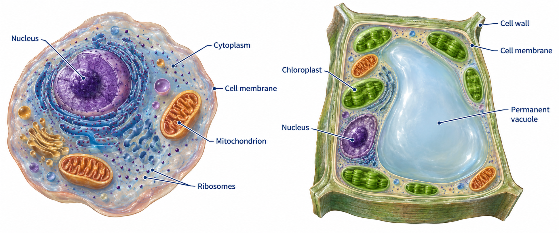
Animal and plant cells are eukaryotic. Their main genetic material is enclosed in a nucleus. Most have these structures:
Nucleus: contains genetic material and controls the cell’s activities.
Cytoplasm: a jelly-like material where many chemical reactions happen. Enzymes control these reactions.
Cell membrane: controls which substances enter and leave.
Mitochondria: where most stages of aerobic respiration happen. Respiration releases energy for cell activities.
Ribosomes: where proteins are made.
Plant cells also have a cellulose cell wall that strengthens the cell. Many have a large permanent vacuole filled with cell sap. Water in the vacuole helps keep the cell firm. Cells in green parts of plants contain chloroplasts. Chlorophyll inside them absorbs light for photosynthesis.
Not every plant cell contains chloroplasts. Root hair cells grow underground and usually have none. A cell wall is outside the cell membrane; it does not replace it.
Compare the shared structures first, then identify the additional plant-cell structures. Diagrams are models, not photographs.
2. Bacterial cells
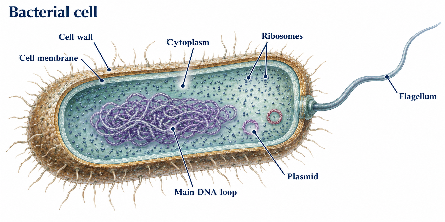
Bacteria are prokaryotic. Their cells are usually much smaller than animal or plant cells. They have cytoplasm, ribosomes and a cell membrane, surrounded by a cell wall. Their wall is not made of cellulose.
Bacteria have no nucleus. Their main genetic material is a single loop of DNA in the cytoplasm. Some also have one or more plasmids, which are small extra rings of DNA. Bacteria do not contain mitochondria or chloroplasts.
“No nucleus” does not mean “no DNA”. Bacteria still have genetic information.
The DNA is not enclosed inside a nucleus. Plasmids are separate from the main DNA loop.
3. Seeing cells: size, magnification and resolution
Cell measurements often use micrometres (µm). 1 cm = 10 mm; 1 mm = 1,000 µm; 1 µm = 1,000 nm. Convert measurements to the same unit before calculating magnification.
Magnification = image size ÷ real size. Rearranging gives real size = image size ÷ magnification. Magnification is a ratio, so an answer such as ×400 has no length unit.
Worked example: calculate a cell’s real size
An image is 24 mm wide at ×600. Real width = 24 ÷ 600 = 0.040 mm. Multiply by 1,000 to convert: 40 µm.
Resolution means telling two close points apart. Simply enlarging a blurred image does not reveal extra detail. Electron microscopes have greater magnification and resolving power than light microscopes, so they reveal much smaller sub-cellular structures.
Standard form uses a number from 1 up to, but not including, 10 multiplied by a power of ten. For example, 0.002 mm = 2 × 10⁻³ mm. A tenfold size difference is one order of magnitude; a hundredfold difference is two.
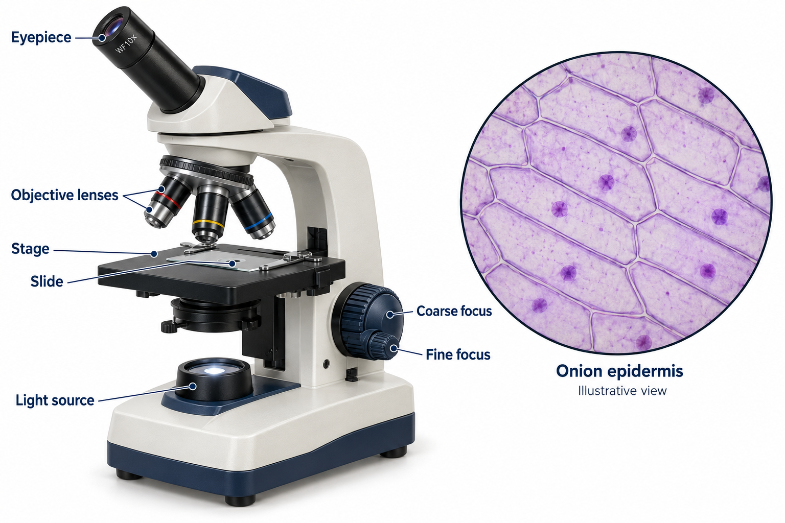
The onion epidermis view is an illustration, not a measured micrograph. For your own drawings, use a calibrated scale bar or calculated drawing magnification.
Required practical 1: light microscopy
Use a light microscope to observe, draw and label plant and animal cells. Use teacher-prepared animal-cell slides or an approved school procedure. Follow instructions for stains, glass slides and biological material.
For a plant specimen, place a thin layer of tissue, such as onion epidermis, in a drop of water on a slide. Add an appropriate stain if instructed.
Lower a coverslip at an angle to reduce trapped air bubbles.
Secure the slide on the stage. Begin with the lowest-power objective and suitable illumination.
Focus with the coarse adjustment, then fine adjustment. Centre the specimen before increasing magnification; use fine focus at high power.
Draw visible structures with clear, single lines. Do not shade. Add straight label lines and a suitable scale bar or calculated drawing magnification.
Total microscope magnification = eyepiece magnification × objective magnification. A ×10 eyepiece and ×40 objective give ×400. A light microscope will not normally resolve individual ribosomes.
4. Specialised cells: structure fits function
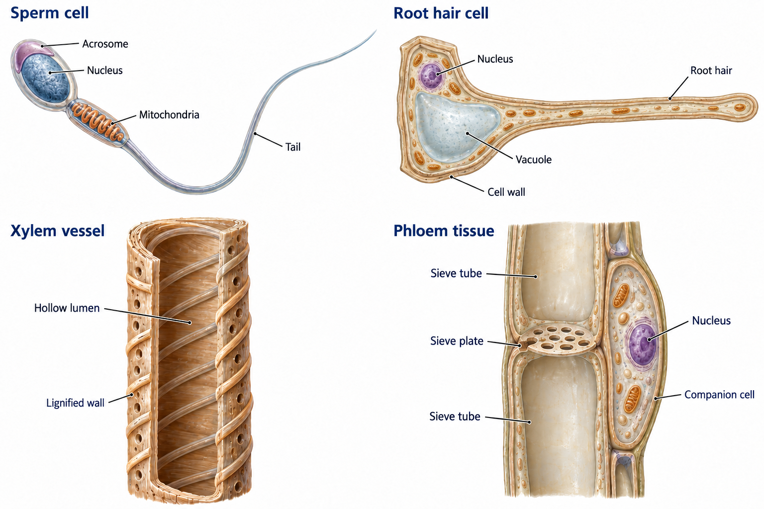
A specialised cell has features that help it do a particular job. To explain an adaptation, link the feature → what it allows → the cell’s job.
Sperm cell: a tail allows movement; many mitochondria release energy for swimming. Enzymes in the acrosome help the sperm enter the egg. Its nucleus carries genetic material.
Nerve cell: a long fibre carries electrical impulses over a distance. Branched ends connect to other cells.
Muscle cell: protein fibres allow contraction. Many mitochondria release energy for contraction.
Root hair cell: its long extension increases surface area for absorbing water and mineral ions. Mitochondria provide energy for active transport of ions.
Xylem vessel: dead, hollow cells join into tubes with no end walls, allowing water flow. Lignin strengthens the walls and helps support the plant.
Phloem: living sieve-tube cells have pores in their end walls, allowing dissolved sugars to pass through. Companion cells support the sieve tubes and provide energy for transport processes.
For each structure, practise finishing the sentence: “This helps the cell because…”
5. Differentiation and the cell cycle
Differentiation happens when a cell develops structures that specialise it for a job. Most animal cell types differentiate early in development. Many plant cells can differentiate throughout the plant’s life. In adult animals, cell division mainly replaces worn-out cells and repairs tissues.
The nucleus contains chromosomes made of DNA. Each chromosome carries many genes. In normal body cells, chromosomes occur in pairs.
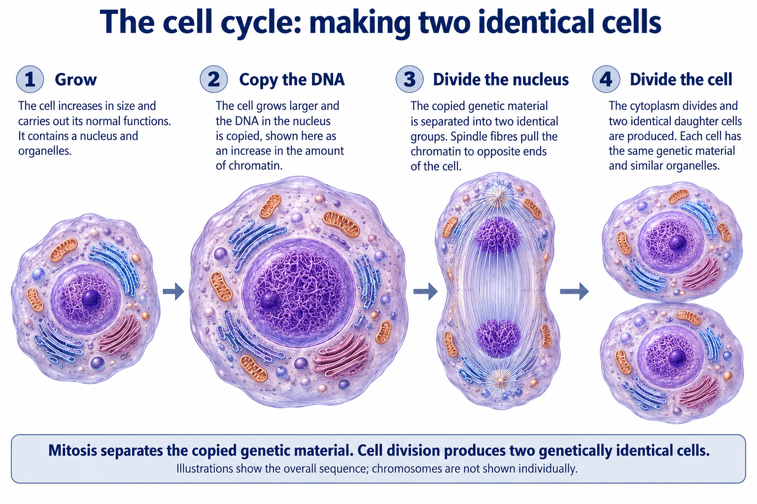
Grow and copy: the cell grows and makes more sub-cellular structures. Its DNA is copied, producing two copies of each chromosome.
Mitosis: the chromosome copies separate into two matching sets at opposite ends of the cell. The nucleus divides.
Cell division: the cytoplasm and cell membrane divide, producing two genetically identical daughter cells.
This is important for growth, development and repair. The daughter cells normally have the same chromosome number as the parent cell. The individual named phases of mitosis are not required here.
Copy the DNA before dividing it. Each daughter cell receives a complete matching set of chromosomes.
6. Stem cells and their uses
A stem cell has not yet specialised. It can divide to make more stem cells and can differentiate into other cell types.
Early human embryos: stem cells can develop into most human cell types.
Adult bone marrow: stem cells can produce a more limited range, including different blood-cell types.
Plant meristems: growing regions contain stem cells that can produce any type of plant cell throughout the plant’s life.
Stem cells could replace damaged cells, for example insulin-producing cells in diabetes or nerve cells in paralysis. A possible use is not the same as a proven, safe treatment. Risks include infection and uncontrolled cell division. Some people object to obtaining cells from embryos on ethical or religious grounds.
In therapeutic cloning, an embryo is produced with the same genes as a patient. Cells derived from it match the patient genetically, reducing the risk of immune rejection. You do not need the laboratory techniques for this course.
Meristem cells can be used to produce clones: genetically identical plants. This can help conserve rare plants and quickly produce many crops with useful features such as disease resistance.
7. Diffusion: moving down a concentration gradient
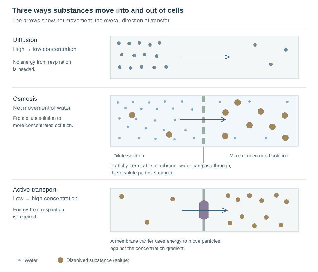
Particles move randomly. When a substance is more concentrated in one place than another, more particles move from the high-concentration side to the low-concentration side than in the reverse direction. This overall movement is diffusion.
Diffusion does not need energy from respiration. Oxygen diffuses into the blood at the lungs; carbon dioxide diffuses out. Urea can diffuse from cells into blood plasma to be carried to the kidneys for removal.
A steeper concentration gradient increases the rate of net movement.
A higher temperature means particles have more kinetic energy and move faster.
A larger surface area allows more particles to cross at once.
A shorter diffusion distance lets particles cross more quickly.
At equal concentrations, particles still move in both directions. There is no net movement: neither side gains overall.
Explore: Diffusion
Particles move randomly. Overall, they spread from a higher concentration to a lower concentration until evenly distributed. They keep moving at equilibrium.
Model limitations: This is a two-dimensional model with very few particles. Their colours, sizes and speeds are not realistic; solvent particles are omitted.
8. Why large organisms need exchange surfaces
A small single-celled organism has a large surface area to volume ratio. Its surface can exchange enough material to supply its small volume. As an organism gets larger, its volume increases faster than its surface area. Diffusion distances also increase.
Larger organisms therefore need specialised exchange surfaces, often linked to transport systems. Effective exchange surfaces have a large area and a short diffusion distance. In animals, blood flow helps maintain concentration gradients. Ventilation replaces air or water at gas-exchange surfaces.
Lungs: many thin-walled alveoli provide a large surface area; blood flow and breathing maintain gradients.
Small intestine: villi provide a large surface area; a thin lining and blood supply help absorb nutrients.
Fish gills: many thin filaments and lamellae give a large area; flowing water and blood maintain gradients.
Roots: root hairs increase the area for absorbing water and mineral ions.
Leaves: broad surfaces, thin tissues, stomata and internal air spaces allow efficient gas exchange.
Worked example: surface area to volume ratio
A cube with sides of 1 mm has surface area 6 × 1² = 6 mm² and volume 1³ = 1 mm³. Its ratio is 6:1. A cube with sides of 2 mm has area 24 mm² and volume 8 mm³, giving 3:1. The larger cube has less surface area for each unit of volume.
9. Osmosis: movement of water
Osmosis is the net movement of water from a dilute solution to a more concentrated solution through a partially permeable membrane. The membrane allows water through but prevents some dissolved substances from crossing.
If potato cells are placed in pure water, water enters the cells and the tissue usually gains mass. In a sufficiently concentrated sugar solution, water leaves the cells and the tissue loses mass. Osmosis does not require energy from respiration.
The moving substance in osmosis is water, not sugar or salt. State which way the water moves and describe the membrane.
Explore: Osmosis
Water crosses a partially permeable membrane in both directions. Initially, more water moves from the dilute solution towards the more concentrated solution. Larger solute particles cannot cross.
Model limitations: This illustrates the initial direction of net movement, not a prediction of final water levels or cell size. Real water movement depends on water potential; pressure and volume changes are not modelled.
Compare the substance that moves, the direction of net movement and whether respiration supplies energy.
Required practical 2: potato tissue and osmosis
Prepare a range of labelled sugar or salt solutions, including pure water.
Cut equal-sized potato cylinders using the same potato type. Use cutting tools only as instructed, on a suitable surface.
Blot and record each cylinder’s initial mass. Put cylinders into equal volumes of solution.
Keep the temperature and time in solution the same. Use repeats for every concentration.
Remove each cylinder, blot it consistently and measure its final mass.
Calculate percentage mass change. Calculate means from repeats and investigate anomalous results.
Percentage change = (final mass − initial mass) ÷ initial mass × 100. A positive value is a gain; a negative value is a loss. Percentages allow comparison when starting masses differ slightly.
Plot solution concentration on the horizontal axis and mean percentage mass change on the vertical axis. Use an appropriate line or curve of best fit. Where it crosses 0% mass change, there was no net water movement. This estimates a solution concentration that balances water movement for that tissue.
Worked example: mass loss and rate
A cylinder changes from 2.50 g to 2.25 g. Change = −0.25 g. Percentage change = −0.25 ÷ 2.50 × 100 = −10%. If this happened over 50 minutes, mean rate of mass loss = 0.25 ÷ 50 = 0.005 g/min.
10. Active transport: against the gradient
Active transport moves substances from a lower concentration to a higher concentration across a membrane. This goes against the concentration gradient and requires energy released by respiration.
Root hair cells use it to take mineral ions from dilute soil water even when the concentration inside the root is higher. Cells in the intestine can absorb sugar into the blood even when its concentration in the gut is lower.
Diffusion and osmosis can occur without energy from respiration. Active transport depends on a supply of this energy, so anything that reduces respiration can reduce active transport.
Explore: Active transport
A carrier protein uses energy supplied by respiration, transferred by ATP, to move particles from lower to higher concentration. Switching off the energy supply stops the pump.
Model limitations: The carrier movement and particle numbers are simplified. Real membrane proteins change shape; the model does not show respiration or the chemical reactions involving ATP.
Scope checked against AQA Combined Science: Trilogy 8464, section 4.1. Original teaching explanations and practice questions; not produced or endorsed by AQA. View the specification.
Put it into practice
02 / 03
Start with the 28 core questions. You can complete this topic over several sessions; your answers save as you go. Use a hint if you get stuck, then compare your work with an example answer. Your tutor can mark written answers.
Question words State / name: give a short answer. Describe: say what happens. Explain: give a reason using “because”. Calculate: show your working and include units.
Diagram, data and practical tasks
Each daughter cell needs a complete set of genetic information.
Build the cell-cycle sequence
Choose a stage for each position, from first to last. The diagram above is there to help you.
Growth and DNA copying both happen before mitosis. The four boxes break the process into smaller learning steps.
Microscopy calculation
A cell image is 18 mm wide. The image has a magnification of ×600. Calculate the real cell width in micrometres (µm).
Step 1: real size = image size ÷ magnification. Step 2: calculate 18 ÷ 600 to get the real size in mm. Step 3: multiply by 1,000 to convert mm to µm.
Read an osmosis result
Equal-sized potato cylinders were placed in different sugar solutions for the same time. The table shows mean results from three cylinders at each concentration. These are invented practice data.
Effect of sugar concentration on potato mass
Sugar / mol/dm³
Mean mass change / %
0.0
+12
0.2
+6
0.4
0
0.6
−6
0.8
−12
Show an example answer
At 0.0 mol/dm³, water moves into the potato cells from the more dilute solution outside, so mass increases. At 0.8 mol/dm³, the solution outside is more concentrated, so water moves out and mass decreases. Water crosses partially permeable cell membranes by osmosis.
On a graph, put concentration on the horizontal axis and percentage mass change on the vertical axis. The 0% crossing here is 0.4 mol/dm³. Water still moves both ways there, but neither side gains overall.
Practice questions
See how far you’ve come
03 / 03
Your answer sheet
When connected with your pupil code, your answers save online for Kevin to see. Click Submit to Kevin when you have finished. Downloads are optional.
Connect with your pupil code above to submit.
One final thought
Show an example answer
For example, a root hair increases the surface area for absorbing water. Water is needed for photosynthesis and helps support plant cells.